ESTHER KUEHN NEUROSCIENCE
  • Overview
    • Publications
    • Lab news
    • MCB >
      • MCB-2017
      • MCB-2019
    • Neuroscience for you >
      • Blog
      • Radio
      • TV_Print
    • Collaborations
    • About me
  • Open positions
  • Research
    • Neuronal Mechanisms
    • Aging and Neurodegeneration
    • MentalHealth
    • BodyMemory
    • Brain in depth >
      • BID-2021 >
        • BID-2021-workshops
        • BID-2021-poster
        • BID-2021-art
        • BID-2021-impressions
      • BID-2019
      • BID-2018
      • BID-2017
    • The team >
      • Publications Claudia Liu
      • Publications Christoph Knoll
      • Publications Juliane Döhler
      • Publications Alicia Northall
      • Publications Avinash Kalyani
      • Publications Kasia Myga
      • Publications Lisa Klemm
      • Publications Jan Oltmer
  • Contact
Picture
My research group applies translational approaches to understand how cortical microstructure links to human brain function in health and disease. We study healthy younger and older adults, people with neurodegenerative diseases and people with mental disorders to understand the neuronal mechanisms that underlie healthy and pathological brain states and their modification. If you would like to join the team contact me.

overview research topics

Picture
Picture
Picture


lab research at a glance

RESEARCH
The organizational principles of de-differentiated cortical maps
A common model assumes that more 'de-differentiated' cortical maps link to worse behavior. Here, we use 7 Tesla MRI to show that cortical de-differentiation is not the common characteristic of older adults' sensory maps, and also does not always link to worse behavior. Read the full eLife paper: [link]
Picture

TREND
Model the human cortex in 3D
We explain why modeling the human cortex in three dimensions, that is, across the cortical surface and in depth, allows novel and unprecedented insights into brain function in health and disease. Key message: In different cortical layers, different computations take place. 3D models of human cognition allow to understand human brain function in its full complexity. Read the full Trends in Cognitive Sciences paper: [link] Read the associated Nature Reviews Neuroscience Commentary: [link] 
Picture

​+++ IN THE NEWS +++
​
​Körpergedächtnis - Negative Gefühle mit positiven bekämpfen


Wie wir weniger grübeln

Wie Körper und Geist zusammen hängen

​
Wenn sich das Gehirn selbst zerstört
​

Warum Altern glücklich macht
​

Eingebrannt ins Gehirn
​

Die Gene sind nicht alles - Was wir gegen Alzheimer tun können
​
Ohnmacht - über das Gefühl, das Leben nicht im Griff zu haben
​

Das Gehirn kann nicht abschalten - Was tun?
​

Posteingang(10.098) - Lesen Sie die Signale, die Ihr Körper Ihnen sendet?
Picture

METHOD
A new computational framework for ultra-high field fMRI
Lack of statistical power and inflated false positive rates have recently been identified as major problems for fMRI analyses. Here, we introduce a novel non-parametric and threshold-free software package called LISA to address this demand. LISA uses a non-linear filter for incorporating spatial context without sacrificing spatial precision. Compared to widely used other methods (e.g., SPM, FLS), LISA boosts statistical power and increases the reliable detection of small activation areas. Key application: The spatial sensitivity of LISA makes it especially suitable for the analysis of fMRI data acquired at ultrahigh field (≥7 Tesla). Read the full Nature Communications paper: [link] 
Picture

KEY-FINDING
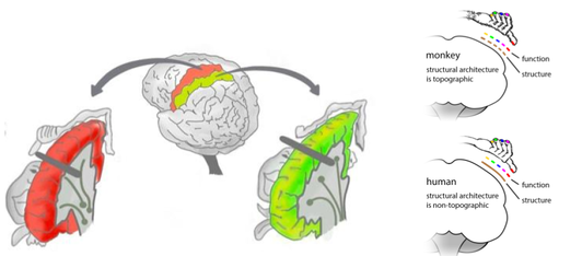
Non-afferent topographic maps in human SI
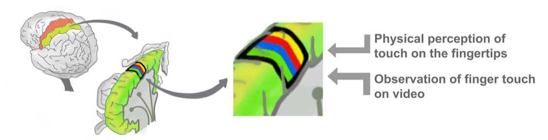
The human primary somatosensory cortex (SI) area 3b has long been believed to code self-perceived (afferent) touch only. We show using 7 Tesla fMRI that feeling touch on the hand and observing touch at another person's hand activates similar fine-grained topographic maps in area 3b and similar inhibitory receptive field interactions. Key insight: Topographic maps in area 3b are also triggered from non-afferent sources. Read the full Journal of Neuroscience and Brain Structure and Function papers: [link] [link]
Picture

KEY-FINDING
Septa in the human brain
Monkeys' and rodents' somatosensory cortices are equipped with myelin-poor septa located in input layer 4 that separate adjacent body part representations. We showed using 7 Tesla MRI that similar boundaries also exist in the human brain.  Human septa are most pronounced in input and output layers of MI, and in input layers of SI. Key insight: Cortical myelin boundaries separate body part representations in the human brain. Read the full Cerebral Cortex paper: [link]
Picture

Powered by Create your own unique website with customizable templates.
  • Overview
    • Publications
    • Lab news
    • MCB >
      • MCB-2017
      • MCB-2019
    • Neuroscience for you >
      • Blog
      • Radio
      • TV_Print
    • Collaborations
    • About me
  • Open positions
  • Research
    • Neuronal Mechanisms
    • Aging and Neurodegeneration
    • MentalHealth
    • BodyMemory
    • Brain in depth >
      • BID-2021 >
        • BID-2021-workshops
        • BID-2021-poster
        • BID-2021-art
        • BID-2021-impressions
      • BID-2019
      • BID-2018
      • BID-2017
    • The team >
      • Publications Claudia Liu
      • Publications Christoph Knoll
      • Publications Juliane Döhler
      • Publications Alicia Northall
      • Publications Avinash Kalyani
      • Publications Kasia Myga
      • Publications Lisa Klemm
      • Publications Jan Oltmer
  • Contact